畜牧与饲料科学 ›› 2023, Vol. 44 ›› Issue (3): 122-128.doi: 10.12160/j.issn.1672-5190.2023.03.018

• 动物疾病防控 • 上一篇    

新疆阿克苏地区蜱和羊感染无浆体的分子流行病学调查

刘凯强1,俞进2,段真真2,金敏1,李佳2,巴音查汗·盖力克1   

  1. 1.新疆农业大学动物医学学院,新疆 乌鲁木齐 830052
    2.新疆维吾尔自治区阿克苏市动物疫病控制诊断中心,新疆 阿克苏 843000
  • 收稿日期:2023-03-01 出版日期:2023-05-30 发布日期:2023-07-12
  • 通讯作者: 李佳(1989—),男,高级兽医师,硕士,主要研究方向为动物疫病监测与流行病学调查。巴音查汗·盖力克(1964—),女,教授,博士生导师,主要研究方向为预防兽医学。
  • 作者简介:刘凯强(1996—),男,硕士研究生,主要研究方向为预防兽医学。
  • 基金资助:
    新疆维吾尔自治区区域协同创新专项-上海合作组织科技伙伴计划及国际科技合作计划项目(2021E01001);新疆维吾尔自治区自然科学基金项目(2022D01F89)

Molecular Epidemiological Investigation of Anaplasma Infection in Ticks and Sheep in Aksu Prefecture,Xinjiang Uygur Autonomous Region

LIU Kaiqiang1,YU Jin2,DUAN Zhenzhen2,JIN Min1,LI Jia2,Bayinchahan Gailike1   

  1. 1. College of Veterinary Medicine,Xinjiang Agricultural University,Urumqi 830052,China
    2. Animal Epidemic Disease Control and Diagnosis Center of Aksu City of Xinjiang Uygur Autonomous Region,Aksu 843000,China
  • Received:2023-03-01 Online:2023-05-30 Published:2023-07-12

摘要:

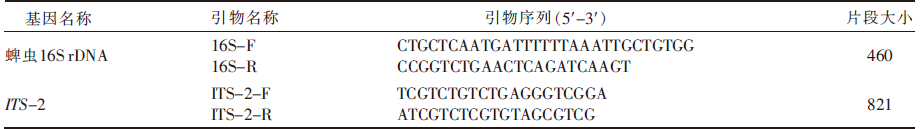
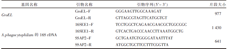
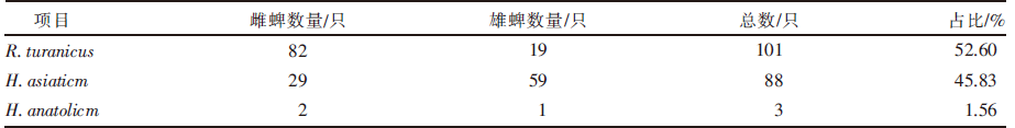
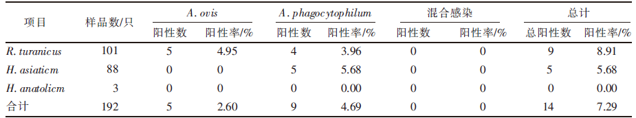
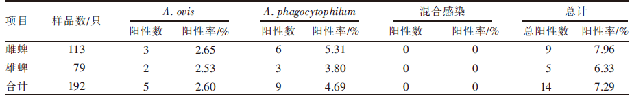
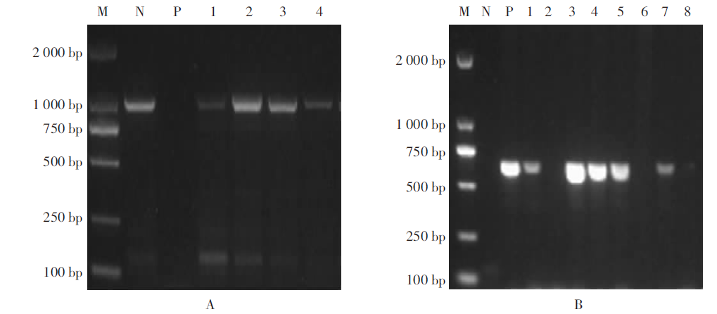
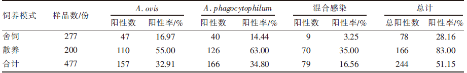
[目的]了解新疆阿克苏地区蜱与羊感染无浆体的情况。[方法]共采集192只蜱和477份羊抗凝血样品;以线粒体16S rDNA和ITS-2为靶基因,采用PCR法对经过形态学鉴定的蜱虫进行分子生物学鉴定;利用特异性引物,分别采用普通PCR法及巢式PCR法检测绵羊无浆体(Ananlasma ovisA. ovis)和嗜吞噬细胞无浆体(Ananlasma phagocytophilumA. phagocytophilum)在蜱和羊抗凝血样品中的携带情况;采用统计学方法分析不同种类、性别蜱虫和不同区域、不同饲养模式下羊感染上述2种无浆体的情况。[结果]采集到的192只蜱虫包括雄蜱79只、雌蜱113只,共有2属3种,分别为扇头蜱属的图兰扇头蜱(Rhipicephalus turanicusR. turanicus)以及璃眼蜱属的亚洲璃眼蜱(Hyalomma asiaticmH. asiaticm)和小亚璃眼蜱(Hyalomma anatolicmH. anatolicm),所占比例分别为52.60%、45.83%、1.56%。蜱样品中,感染2种无浆体的总体阳性率为7.29%,未检测到A. ovisA. phagocytophilum混合感染;R. turanicusH. asiaticmH. anatolicm感染无浆体的阳性率分别为8.91%、5.68%、0,三者差异有统计学意义(χ2=3.20,P<0.05);雄蜱和雌蜱感染无浆体的阳性率分别为6.33%、7.96%,二者差异无统计学意义(χ2=0.30,P>0.05)。羊抗凝血样品中,感染2种无浆体的总体阳性率为51.15%,与蜱样品的总体阳性率差异有统计学意义(χ2=111.35,P<0.05);A. ovisA. phagocytophilum混合感染率为16.56%;舍饲羊和散养羊感染无浆体阳性率分别为28.16%和83.00%,二者差异有统计学意义(χ2=139.81,P<0.05);库车市和沙雅县羊无浆体阳性率最低,为5.00%,温宿县阳性率最高,为64.83%,二者差异有统计学意义(χ2=27.72,P<0.05)。[结论]阿克苏地区蜱和羊普遍感染无浆体,应该加强对无浆体病的防治工作。

关键词: 蜱, 种属鉴定, 绵羊无浆体, 嗜吞噬细胞无浆体, 分子流行病学调查

Abstract:

[Objective] The aim of this study was to understand the prevalence of Anaplasma infection in ticks and sheep in Aksu Prefecture of Xinjiang Uygur Autonomous Region. [Method] A total of 192 ticks and 477 sheep anticoagulant blood samples were collected. After morphological identification, the ticks were molecularly confirmed by using PCR assays targeting mitochondrial 16S rDNA and ITS-2 genes. The presence of Anaplasma ovisA. ovis) and Ananlasma phagocytophilumA. phagocytophilum) in the ticks and sheep anticoagulant blood samples were detected by using conventional PCR assay and nested PCR assay with specific primers, respectively. Statistical analysis were performed to assess the the differences in prevalence of A. ovis and A. phagocytophilum among ticks of different genders as well as sheep reared in various regions and feeding modes. [Result] The 192 ticks, including 79 male ticks and 113 female ticks, belonged to 2 genera and 3 species. The number of ticks identified as Rhipicephalus turanicusR. turanicus, Hyalomma asiaticmH. asiaticm) and Hyalomma anatolicumH. anatolicum) accounted for 52.60%, 45.83% and 1.56% of the total, respectively. In the tick samples, the overall positive rate of Anaplasma infection was 7.29%, while the mixed infection of A. ovis and A. phagocytophilum was not observed; the positive rates of Anaplasma infection for R. turanicus, H. asiaticm, and H. anatolicm were 8.91%, 5.68%, and 0, respectively, with statistically significant differences (χ2=3.20, P<0.05); the positive rates of Anaplasma infection for male and female ticks were 6.33% and 7.96%, respectively, with no statistically significant difference (χ2=0.30, P>0.05). In the sheep anticoagulant blood samples, the overall positive rate of Anaplasma infection was 51.15%, which had statistically significant difference with that of the tick samples (χ2=111.35, P<0.05); the mixed infection rate of A. ovis and A. phagocytophilum was 16.56%; the positive rates of Anaplasma infection in captive and free range sheep were 28.16% and 83.00%, respectively, with statistically significant difference (χ2=139.81, P<0.05); Kuqa City and Xayar County had the lowest positive rate of Anaplasma infection at 5.00%, and Wensu County had the highest positive rate at 64.83%, with statistically significant difference (χ2=27.72, P<0.05). [Conclusion] The prevalence of Anaplasma infection in ticks and sheep in Aksu Prefecture was common, and the prevention and control of anaplasmosis should be strengthened.

Key words: tick, species identification, Anaplasma ovis, Ananlasma phagocytophilum, molecular epidemiological investigation

中图分类号: