畜牧与饲料科学 ›› 2023, Vol. 44 ›› Issue (4): 17-21.doi: 10.12160/j.issn.1672-5190.2023.04.003

• 基础研究 • 上一篇    下一篇

Caspase-9蛋白在亚慢性砷染毒大鼠睾丸间质细胞中的表达研究

秦海霞1,韩菲1,戴研平2,刘改萍1   

  1. 1.乌兰察布医学高等专科学校组胚教研室,内蒙古 乌兰察布 012000
    2.岳阳职业技术学院临床医学院,湖南 岳阳 414000
  • 收稿日期:2023-05-24 出版日期:2023-07-30 发布日期:2023-08-30
  • 通讯作者: 韩菲(1987—),女,副教授,硕士,主要从事生殖学研究工作。
  • 作者简介:秦海霞(1976—),女,讲师,硕士,主要从事生殖学研究工作。
  • 基金资助:
    内蒙古自治区高等学校科学研究项目“砷对大鼠睾丸组织水通道蛋白-7、Survivin影响的研究”(NJZY19335)

Expression of Caspase-9 Protein in Leydig Cells of Rats with Sub-chronic Arsenic Poisoning

QIN Haixia1,HAN Fei1,DAI Yanping2,LIU Gaiping1   

  1. 1. Department of Histology and Embryology,Ulanqab Medical College,Ulanqab 012000,China
    2. Department of Clinical Medicine,Yueyang Vocational and Technical College,Yueyang 414000,China
  • Received:2023-05-24 Online:2023-07-30 Published:2023-08-30

摘要:

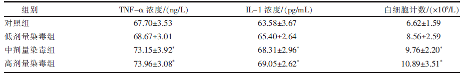
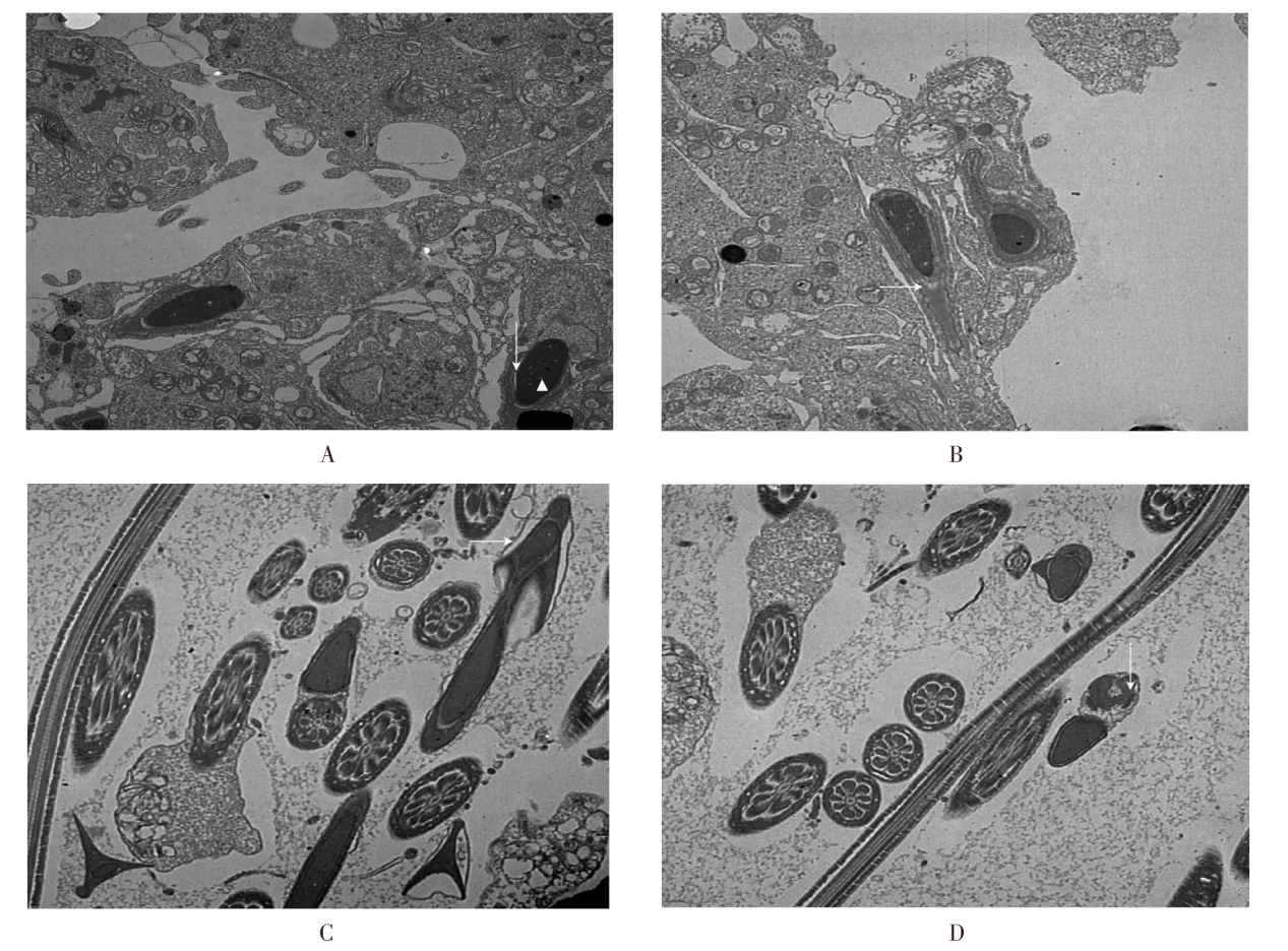
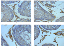
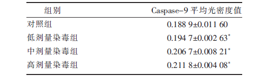
[目的]探讨亚慢性砷染毒对大鼠睾丸间质细胞Caspase-9蛋白表达的影响。[方法]将40只健康成年清洁级SD雄性大鼠随机分为4组,分别为对照组以及低、中、高剂量染毒组,每组10只。采用自由饮水方式进行染毒,低、中、高剂量染毒组亚砷酸钠的染毒剂量分别为2.4、12、60 mg/L,对照组给予生理盐水,染毒试验持续14周。染毒试验结束后,检测大鼠外周血白细胞数量;采用ELISA法检测大鼠血清TNF-α及IL-1的浓度;利用透射电镜观察大鼠精子的超微结构;应用免疫组化法检测大鼠睾丸间质细胞Caspase-9蛋白的表达定位及表达水平。[结果]与对照组相比,中、高剂量染毒组大鼠血清中TNF-α、IL-1浓度及白细胞计数均显著(P<0.05)升高。透射电镜下,对照组精子顶体结构完整,厚度及染色均匀;低剂量染毒组可见精子胞膜肿胀,核染色质较均匀;中剂量染毒组精子顶体与核分离,胞膜肿胀;高剂量染毒组精子尾部中断,轴丝排列紊乱。免疫组化法分析表明,Caspase-9蛋白主要表达于睾丸间质细胞的细胞质,表达信号呈棕褐色;与对照组相比,各剂量染毒组大鼠睾丸组织内Caspase-9蛋白的表达水平均显著(P<0.05)升高。[结论]睾丸间质细胞Caspase-9蛋白表达水平升高可能是亚砷酸钠致雄性大鼠慢性生殖毒性的机制之一。

关键词: 亚慢性砷中毒, 睾丸间质细胞, 超微结构, Caspase-9蛋白, 大鼠

Abstract:

[Objective] This study was conducted to investigate the effects of sub-chronic arsenic poisoning on the expression of Caspase-9 protein in leydig cells of rats. [Method] A total of 40 healthy adult clean-grade male SD rats were randomly assigned into the following 4 groups:a control group and the low-, medium-, and high-dose arsenic exposure groups, with 10 rats in each group. The rats in the low-, medium-, and high-dose arsenic exposure groups were given sodium arsenate by drinking water at concentrations of 2.4, 12 and 60 mg/L, respectively, and those in the control group was given physiological saline. The exposure test lasted for 14 weeks. Following the exposure test, the number of peripheral white blood cells of the rats was counted; the serum levels of TNF-α and IL-1 were measured by ELISA assay; the ultrastructure of sperm was observed using transmission electron microscopy; the expression level and localization of Caspase-9 protein in leydig cells were assessed by immunohistochemical method. [Result] Compared with the control group, the serum levels of TNF-α and IL-1 as well as the number of peripheral white blood cells in the medium- and high-dose arsenic exposure groups significantly (P<0.05) increased. Under transmission electron microscopy, the sperm acrosome structure in the control group was intact, with uniform thickness and staining. In the low-dose arsenic exposure group, the sperm membrane swelled and the nuclear chromatin was uniform. In the medium-dose arsenic exposure group, the sperm acrosome and nucleus split, and the membrane swelled. In the high-dose arsenic exposure group, the sperm tail disrupted and the axial filament arrangement disordered. Immunohistochemical analysis showed that Caspase-9 protein mainly expressed in the cytoplasm of leydig cells, and the expression signal was dark brown. Compared with the control group, the expression level of Caspase-9 protein in the testicular tissue in all the arsenic exposure groups significantly (P<0.05) increased. [Conclusion] The increased expression level of Caspase-9 protein in laydig cells may be one of the mechanisms underlying chronic reproductive toxicity induced by sodium arsenite in male rats.

Key words: sub-chronic arsenic poisoning, leydig cell, ultrastructure, Caspase-9 protein, rat

中图分类号: